by D. Van Metre 1 (5/2010)
Quick Facts…
- The progression of clinical signs of pinkeye is consistent and predictable.
- Pinkeye typically occurs in an outbreak, affecting first lactation cows soon after calving. Younger and older animals may also be affected but not as frequently.
- The goals of pinkeye treatment are to eliminate the causative agent using antibiotics and to prevent rupture of the eye.
Introduction
Pinkeye is known technically as Infectious Bovine Keratoconjunctivitis, in recognition of its contagious nature and the damage that results to the cornea (the clear portion of the eye) and conjunctiva. The causative agent is Moraxella bovis, but other factors, such as direct sunlight and dust, are thought to be necessary in order for the disease to be present. Flies are important vectors involved in spread of the organism from one cow to another. The immune status of the cow also appears to play an important role in the expression of the disease. Cows are almost never affected with pinkeye more than once in their lifetimes and seldom are affected in more than one eye.
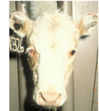 |
| Figure 1: Healing phase of pinkeye with decreased pain and tearing, decreased clouding of the cornea, and early scar formation as the ulcer heals. |
The progression of clinical signs of pinkeye (Figure 1) is consistent and predictable. Excessive tearing, reddening of the conjunctiva, and frequent blinking (lacrimation, conjunctivitis, and blepharospasm) are followed within one to two days by corneal ulceration and cloudiness (edema). The ulcer begins in the center of the cornea and may expand to occupy most of the cornea. In severe cases, the ulcer may erode the entire thickness of the cornea, resulting in rupture of the eye and permanent blindness. Corneal edema may quickly affect the entire cornea, resulting in the characteristic “blue eye” seen in advanced cases. The eye is blind and very painful and the animal will hold it shut and avoid the sun.
Pinkeye typically occurs in an outbreak, affecting first lactation cows soon after calving. Younger and older animals may also be affected but not as frequently. Due in part to differences in risk factors and the presence of herd immunity, outbreaks occur at intervals of three to five years with sporadic isolated cases appearing between outbreaks.
Treatment
The goals of pinkeye treatment are to eliminate the causative agent using antibiotics and to prevent rupture of the eye. Moraxella bovis is susceptible to most antibiotics including penicillin and oxytetracycline. Early cases (tearing and conjunctivitis without ulcer and edema) are successfully treated with topical antibiotics. We recommend that during an outbreak, first lactation cows be examined daily and at the firunk lockups simplify this procedure immensely. Such early topical treatment will usually prevent progression to ulceration. Eyes with corneal ulcers require aggressive antibiotic treatmst sign of tearing, 1-2 cc of penicillin be squirted in the affected eye for two to three days. Feedbent and veterinary assistance should be secured. We prefer the use of subconjunctival injections of 1 cc of penicillin to achieve high levels of antibiotics in the eye. Neither of these uses of penicillin (topical or subconjunctival) will result in violative residues of antibiotics in the milk or meat. In beef cattle, a single dose of long-acting oxytetracycline has been used very successfully in early cases. Similar treatment is now approved in dairy cattle, but withholding of milk will be necessary.
If the corneal ulcer is deep and appears likely to rupture, we recommend the eyelids be sewn shut or the third eyelid sewn to the upper lid across the ulcer. This procedure is best left to a veterinarian. “Pinkeye patches” are quite popular as they are easy to apply. However, their only value is to protect the eye from the sun. Eyes covered with a patch cannot be medicated or monitored for progression of the disease.
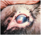 |
| Figure 2: Pinkeye that is several days old shows severe tearing, a blue color to the cornea due to edema, and partially closed eyelid due to pain and sensitivity. |
Healing of corneal ulcers progresses in stages (Figure 2). Once the organism is eliminated, epithelium quickly covers the ulcer and the eye is much less painful. The eye will cease its excessive tearing and blinking and the animal will appear much more comfortable. Even though the “blue-eye” (edema) and blindness may remain for one to two weeks after the ulcer is healed, the eye no longer requires antibiotic treatment. Clearing of the edema begins at the periphery of the cornea and sight will return accordingly. A central scar at the site of the ulcer will gradually shrink over a period of several weeks, but a small opaque area may remain for the lifetime of the animal.
Prevention
Prevention of pinkeye is limited to fly control, since little can be done to reduce dust or exposure to sunlight. Several vaccines are available, most of which stimulate significant levels of circulating antibody against the organism or its pili, which facilitate attachment to the conjunctiva. There are numerous anecdotes in which producers describe the worst outbreak of pinkeye ever seen in their herd. These producers vaccinated their herds the following winter and no pinkeye was seen that summer. However, controlled trials have failed to show that vaccination will reduce the incidence of naturally occurring pinkeye. The apparent benefits of the vaccine seen the year after pinkeye outbreaks are almost certainly due to herd immunity stimulated not by the vaccine but by the outbreak itself.
1 F.Garry, DVM, M, DACVIM, Colorado State University Extension Specialist and professor, (Veterinarian), College of Veterinary Medicine and Biomedical Sciences, Clinical Sciences. (5/2010).
Colorado State University, U.S. Department of Agriculture and Colorado counties cooperating. Extension programs are available to all without discrimination. No endorsement of products mentioned is intended nor is criticism implied of products not mentioned.